The CellAssist
The CellAssist
Benchtop Live Cell Imager
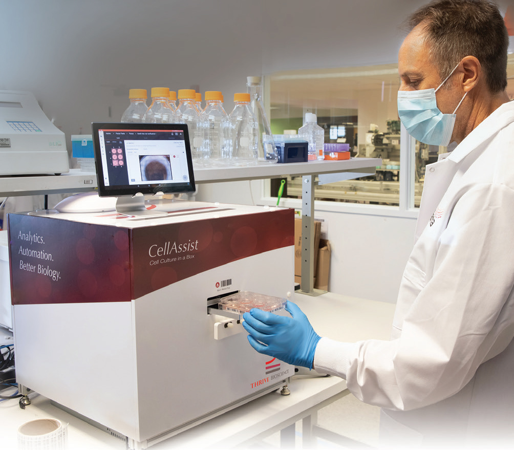
Thrive Bioscience’s new benchtop instrument, the CellAssist, automatically builds a database of images, metrics, and methods, and materials to ensure centralized capture, and retention of data. The unique CellAssist sets new standards for reproducible cell, tissue, and suspension culture in numerous applications.
- The CellAssist replaces your time at the inverted microscope to acquire, organize, and analyze images of all your cells in culture plates.
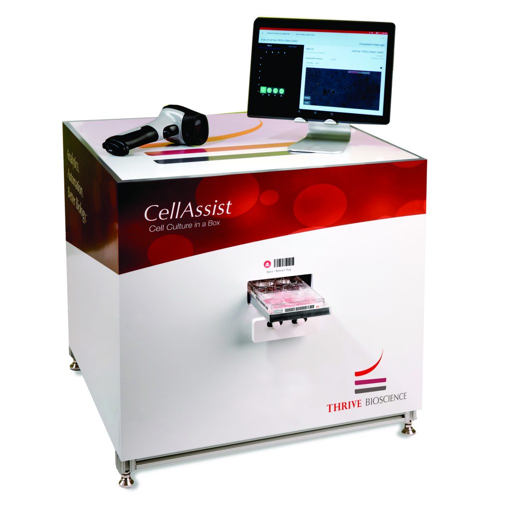
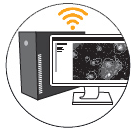
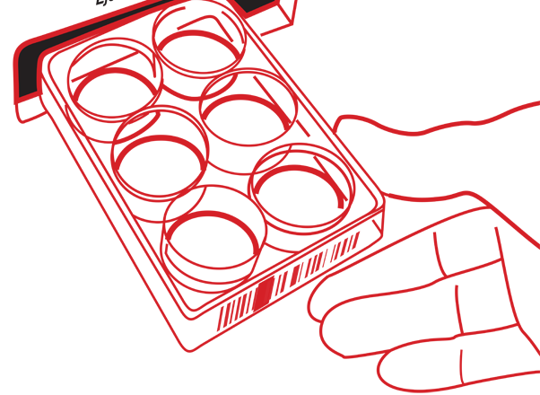
- Minutes after inserting a standard 6-well through 384-well flat bottom or round bottom plate into the CellAssist, the system captures, displays, and processes hundreds of high-resolution images of adherent cells, suspension cells, and organoids at 4x, 10x, and 20x, using phase contrast or bright-field imaging.
- The CellAssist captures images at 100+ focal planes, each 2 μm to 50 μm apart (user selectable), with a z-range of 4.0 mm
- With barcodes to identify plates, and reagents, the CellAssist maintains a complete centralized record of growth, and behavior of all cells.
CellAssist Software
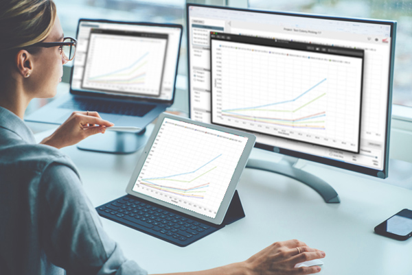
Thrive Bioscience’s CellAssist Software is a computer workstation at the heart of the CellAssist’s visualization and analysis capabilities. It can be placed in the laboratory or office to automatically download all of the images from the CellAssist instrument.
The CellAssist software analyzes the images, and presents a comprehensive, and coherent view into all of the images, and metrics from the entire history of all of the laboratory’s projects.
In addition, the entire plate setup information, normally entered into the CellAssist tablet, can also be entered into CellAssist Software, and automatically transmitted to the CellAssist instrument.
CellAssist Features, and Specifications
The CellAssist provides image capture, reports, and a database on cultured cells with these added features, and product specifications:
Key Features
Instrumentation
- Barcode tracking of culture plates, reagents, and workflow
- Imaging of adherent cells, suspension cells, and organoids in standard 6-well through 384-well plates
- Scanning modes include center-of-well, standard-well, whole-well, and regions of interest
- Wireless, and wired network connection between CellAssist instrument(s), and EvalCore Analysis Workstation, enabling location installation flexibility
Software
- Central database of images, and metrics, including growth rates, confluence, colony count, and size, and monoclonality
- Detailed viewing of stitched images at varying z-height depths along with zoom, measurement, and export capabilities
- Time lapse viewing capability to visualize colony formation, morphology, differentiation, and monoclonality
Instrument Specifications
- Microscopy: 4x, 10x, and 20x standard; phase contrast or bright-field
- Image Field of View: 5.3mm x 4.0mm at 4x; 2.1mm x 1.6mm at 10x
- Resolution: 2.1 μm/pixel at 4x; 0.84 μm/pixel at 10x/0.41 µm/pixel at 20x
- Focal Planes: Captures images at up to 100 focal planes, 2 μm to 50 μm apart (user selectable), with a z-range of 3.5 mm
- User interfaces: Tablet in laboratory, and EvalCore Analysis Workstation in laboratory or office
- Accessories Included with CellAssist:
- Barcode reader (Bluetooth) for plate ID, reagents, and experiment data entry
- Tablet user interface (Wi-Fi) in laboratory for viewing, and project planning
- Wireless access point for private network with EvalCore Analysis Workstation
- Physical:
- Internal temperature: 37°C with CO2 option
- Local storage: 4 TB
- Dimensions: 0.5m x 0.5m x 0.5m (lwh) / Weight: 32 kg
- Operating environment: temp 20 to 27°C; relative humidity 20 to 80%
- Power requirements: 100 to 240VAC 50/60Hz; 650W max
Instrument Specifications
- Powerful computing workstation combined with CellAssist, and CA Control software for image analysis, and project management
- Networked to the CellAssist with either a wired or wireless connection
- Automatic image transfer from CellAssist instrument(s)
- Image, and video export formats: png, mp4
- Physical:
- Local storage: 18TB
- Dimensions: 0.5m x 0.2m x 0.8m (lwh) (without monitor) / Weight: 14 kg
- Power requirements: 100 to 240VAC, 50/60Hz; 890W max
Instrumentation
- Barcode tracking of culture plates, reagents, and workflow
- Imaging of adherent cells, suspension cells, and organoids in standard 6-well through 384-well flat or round-bottom plates
- Scanning modes include center-of-well, standard-well, whole-well, and regions of interest
- Wireless, and wired network connection between CellAssist instrument(s), and EvalCore Analysis Workstation, enabling location installation flexibility
Software
- Central database of images, and metrics, including growth rates, confluence, colony count, and size, and monoclonality
- Detailed viewing of stitched images at varying z-height depths along with zoom, measurement, and export capabilities
- Time lapse viewing capability to visualize colony formation, morphology, differentiation, and monoclonality
- Microscopy: 4x, 10x, and 20x standard; phase contrast or bright-field
- Image Field of View: 5.3mm x 4.0mm at 4x; 2.1mm x 1.6mm at 10x; 1.1 mm x 0.8 mm at 20x
- Resolution: 2.1 μm/pixel at 4x; 0.84 μm/pixel at 10x; 0.41 µm/pixel at 20x
- Focal Planes: Captures images at 100+ focal planes, 2 μm to 50 μm apart (user selectable), with a z-range of 4.0 mm
- User interfaces: Tablet in laboratory, and EvalCore Analysis Workstation in laboratory or office
- Accessories Included with CellAssist:
- Barcode reader (Bluetooth) for plate ID, reagents, and experiment data entry
- Tablet user interface (Wi-Fi) in laboratory for viewing, and project planning
- Wireless access point for private network with EvalCore Analysis Workstation
- Physical:
- Internal temperature: 37°C with CO2 option
- Local storage: 4+ TB
- Dimensions: 0.5m x 0.5m x 0.5m (lwh) / Weight: 32 kg
- Operating environment: temp 20 to 37°C; relative humidity 20 to 80%
- Power requirements: 100 to 240VAC 50/60Hz; 650W max
- Powerful computing workstation combined with CellAssist, and CA Control software for image analysis, and project management
- Networked to the CellAssist with either a wired or wireless connection
- Automatic image transfer from CellAssist instrument(s)
- Image, and video export formats: png, mp4
- Physical:
- Local storage: 18TB
- Dimensions: 0.5m x 0.2m x 0.8m (lwh) (without monitor) / Weight: 14 kg
- Power requirements: 100 to 240VAC, 50/60Hz; 890W max
Specifications are subject to change without notice. The CellAssist is for in vitro, and laboratory use only.
Video: The CellAssist Experience
“The CellAssist Experience” video (3:38 minutes) provides an overview of the CellAssist, which combines an advanced imaging system coupled with intuitive software to provide high-resolution images with excellent accuracy.
The unique capabilities outlined in the video enable biologists to gain a deeper insight into cell dynamics, and greatly improve the reproducibility of cell, tissue, and suspension culture experiments. Learn more about CellAssist, and its capabilities with this video.
CellAssist Workflow
Video: A Researcher's Insight
Thrive Bioscience recently conducted a comprehensive video interview with Steven Sheridan, PhD, Senior Research Investigator, Center for Genomic Medicine, Massachusetts General Hospital (8:48 minutes).
During this “Researcher’s Insight” interview, Steven Sheridan provided an in depth overview of how the CellAssist addresses his laboratory’s cell culture pain points. Specifically, how to empower biologists with superior image resolution, excellent registration across multiple scans, and a centralized database to capture consistent, and shareable images, methods, and materials to support reproducible cell, tissue, and suspension culture research.
CellAssist Benefits For Your Laboratory
Centralized database provides consistent, and shareable images
to improve your cell, tissue, and suspension culture
experiments, and results
- Organizes images across time, cell lines, and labs
- Images, and displays cells at the same location on the cell, tissue, and suspension culture plate within 15 μm across scans
- Generates extensive data sets of over 3 GB of images per scan
Objective metrics, and quantitative reports
enhance cell, tissue, and suspension culture
decision making, and productivity
- Characterizes cell dynamics, including growth curves, confluence, and the number, and area of colonies
- Analyzes, and compares cells to support decision-making to reduce variability, and increase quality
- Allows comparison, and optimization of multiple methods, and materials
Key workflow details, and status of cells are captured, and archived
to provide confidence that data is not lost or compromised
- Accurately records, and time-stamps your cell, tissue, and suspension culture process with a barcode scanner
- Captures, and archives the key workflow details, and the status of cells to provide confidence that data is not lost or compromised
- Enables comparison with previous experiments to optimize, and ensure reproducibility of your cell, tissue, and suspension culture
